When accuracy matters, Chirascan helps you see what other techniques miss — from pH effects to oxidation-induced fragmentation.
By: Mohammad Shadab
Detecting change
Revealing subtle structural shifts in therapeutic antibodies through high-sensitivity CD.
Detecting change: Can circular dichroism offer greater sensitivity when analyzing changes to therapeutic antibodies?
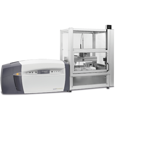
Informed decision-making is pivotal during the analysis, formulation and process development of therapeutic antibodies. Using Higher Order Structure (HOS) comparisons is a key part of this, ensuring accurate biophysical characterization of the biotherapeutic proteins. In order to get a sensitive and precise picture of the way a therapeutic antibody may change under stress conditions; it is important that there is no room for subjectivity in interpreting the spectral data. The Chirascan Q100 technology uses a 96-well microplate format which allows for the setting of clear acceptance criteria and works hard to eliminate needless subjectivity inherent to visual data interpretation.
Greater experimentation, better decision-making
The importance that circular dichroism plays in characterizing the HOS for both biochemical and functional studies was recently recognized by the FDA. Here at Applied Photophysics, we work hard to test and develop the techniques that make this possible. Our recent case study, conducted in collaboration with UCB Pharma and the Centre for Process Innovation (UK), scrutinized the revolutionary Chirascan Q100 technology. Read on to find out how it fared when put to the test.
Case study: A summary
The recent case study collected data from circular dichroism measurements, statistically validated these and then compared them to the results from orthogonal and complementary techniques. This comparison meant that we were provided with a holistic picture of the stability of antibody products. Importantly, the cross-data comparisons show very clearly how different forced degradation conditions impact this stability. The result? A detailed analysis of both secondary and tertiary structures of antibody products that revealed a sensitive picture of the changes these structures undergo when in stress conditions.
The experiment: details and data analysis
- A range of forced degradation conditions was set for 14 days, and monoclonal antibody (Fab and IgG) samples were subjected to these. Dilution of all samples was set to 0.8 mg/mL in 20 mM NaPi, 150 mM NaF, pH 6.
- Flow cells with a pathlength of 0.2 mm were used for far-UV CD experiments, and flow cells with a pathlength of 10 mm were used for near-UV CD experiments.
- Acquisitions were performed at 20°C, 1 s timer-per-point, 1 nm bandwidth, 0.5 nm step size and 3 repeat scans. Each measurement comprised 6 replicates per sample.
Before HOS comparisons, baseline-corrected CD spectra were normalized by absorbance. The baseline correction used an individual buffer reference for each replicate and the final stage comprised the averaging of repeat scans. HOS comparisons were carried out in terms of Weighted Spectral Difference (WSD). If 4 or more out of 6 replicated were outside the quality range, then minor spectral differences were considered statistically significant (having a WSD value of >50%).
The results of our case study: HOS comparisons
The HOS comparisons, following the high- sensitivity CD analysis, showed up some minor changes in the secondary and tertiary structure. This was revealed in some of the stress conditions, particularly light stress, pH, and oxidation. The sensitive nature of our statistical analysis demonstrated that some of these changes statistical significance.
The round-up: Overall conclusions
The most noteworthy conclusion of our case study is that the results from CD techniques are a valuable complement to orthogonal techniques. This combination provides a marked improvement in the sensitivity of data-analysis and can show an impressive range of results.
In particular:
- CD is more sensitive to light stress in near-UV experiments than far-UV and cIEF.
- Although CD is less-sensitive to temperature stress, it is particularly suited to picking up the impact of pH-change on HOS.
- For IgG1, the process of CD is sensitive to fragmentation due to oxidation. This is true for both far- and near-UV CD.
The benefits: What does CD allow?
CD is a great tool for improving decision-making, for ensuring you can make informed decisions about time investment. The quick screening that CD allows for both PTMs and aggregation/fragmentation means it is an important instrument in, for example, down-stream characterization. Furthermore, the automation that CD offers means that reproducibility can increase, and an ample number of replicates can be produced. This automation doesn’t compromise on robust statistical analysis or producing HOS comparisons without user-bias. All of this comes with improved sensitivity to detecting minor changes in the tertiary structure of therapeutic antibodies, due to the solid-state detector.
Where CD Precision Meets Possibility